Lab 1: Principles and use of microscope
SCHOOL OF TECHNOLOGY INDUSTRY
UNIVERSITY SCIENCES MALAYSIA
DEGREE IN BIOPROCESS
IBG 102
BIOLOGY FOR TECHNOLOGIST
| Name | 1. LOH SHI WEI (137602) 2. LAI CHONG SING (137592) 3. SITI NORASYIKIN BINTI SALMI (137673) 4. SITI NUR SUHAILI AFIQAH BINTI SARIMAN (137674) 5. NUR LIYANA ATHILAH BINTI MOHD AFFANDI (137636) |
| Title | 1.1 SETTING UP AND USING THE MICROSCOPE 1.2 EXAMINATION OF CELLS |
| Date of practical | 19/09/17 |
| Date of Report Submitted | 25/09/17 |
| Lecturer | DR. TYE |
LAB 1: PRINCIPLE AND USED OF MICROSCOPE
1.1 Setting Up And Using The Microscope.
INTRODUCTION
A microscope is an equipment which is used to magnify the organisms or the structure which cannot be seen with our naked eyes. There are two categories of microscope, which is light microscope and electron microscope. Light microscope is a microscope which allows the light to pass through the specimen through the objective lens band eyepiece lens. This microscope is suitable to study the living and unstained specimens. Meanwhile, for electron microscope, it uses a beam of electron instead of beam of light to produce a short wavelength. It has higher resolution of power to produce higher magnification of image of the specimen. For light microscope, it can be divided into six sub-categories, which is bright field, dark field, ultraviolet, fluorescence, phase contrast and differential interference contrast (D.I.C)

Figure 1.1 shows the light microscope.
The part of light microscope
- Eyepiece lens: The lens that you look through. It usually has the magnifying power of 10X or 15X, which transmits the light from the objective lens to our eyes.
- Eyepiece tube: Hold the eyepieces in place above the objective lens. It helps to receive the light coming through the objective lens and redirects it to the eyepieces.
- Objective lens: An optical lens to gather and focus the light to magnify an image of the specimen. Usually, they have four types of objective lens with magnification power of 4X, 10X, 40X and 100X. When coupled with the eyepiece e.g. 10X ), It will give the magnification of 40X (4X x 10X), 100X, 400X and 1000X. The longer the objective lens, the greater the power would be.
- Nosepiece : Holds the objective lens and can rotate to change the power easily.
- Stage : The flat platform to support the slide. For mechanical stage, it can adjust the position of slide by using the knobs provided.
- Condenser : It helps to focus the light from the light source onto the specimen. It is useful for attaining the specimen up to 400X and above.
- Iris diaphragm: It confines the amount of light reaching to a specimen. It is located above the stage and below the condenser.
- Coarse focus adjustment knob: It is used to focus on the specimen.
- Fine focus adjustment knob: To control precise focusing on the specimen after using the course adjustment knob.
- Light source bulb: It provides the light source and illumination of the specimen.
- Arm : The support structure that connect the lens system to the base
- Base : It contains the light source and support the structure
Magnification is the ability to enlarge the appearance of the object which is small and tiny. It is better to start with the lower magnification to get specimen into focus, later proceed to a higher magnification. The total magnification of the microscope is the power of the objective lens times with the power of eyepiece lens. There would be four types of magnification in the microscope.
| Power of eyepiece lens | Power of objective lens | Total magnification |
| 10X | 4X | 40X |
| 10X | 10X | 100X |
| 10X | 40X | 400X |
| 10X | 100X | 1000X |
Resolution of microscopy is the ability to differentiate two very small objects from each other. When closing the diaphragm, the light is more confines to the condenser. The contrast of image increase but the resolution power of the image decrease. When opening the diaphragm, more light are going into the condenser. This will decrease the contrast but increase the resolution of the image.
OBJECTIVE
- To improve the skills on handling a simple bright-field microscope
- To learn the important part of the microscope
- To learn the important of magnification and resolution of microscope
- To learn the ways to take care of the microscope with correctly
MATERIALS AND REAGENT
Microscope slide and cover-slip
PROCEDURE
A) Preparation:
- Sat on the stool with knees under the bench and the microscope is moved so that both eyepieces were looked through without straining to ensure own comfortability.
- The power lead of the microscope is plugged in and the power is turned on, the microscope light is turned on by using the main on-off switch.
- The light intensity is adjusted using the brightness control. The normal adequate position is 5.
- The revolving nosepiece is rotated to bring the 4x objective lens into the light path.
- A clean slide is taken and a line is marked on it using a marker pen. The slide is placed on the stage and it is secured by using the spring clip. The slide is moved into the light path using the coaxial stage control knobs.
- Both eyepieces were being looked through and were adjusted until a single circle of light is seen. A note is made in the class manual of the setting on the interpupillary distance scale for future reference.
- The tube length adjustment (diopter) ring on the right eyepiece is rotated to match the interpupillary distance setting obtained in 1.6.
- The marker pen mark is focused by adjusting the coarse and fine adjustment knobs which is done by using only right eye.
- The left eyepiece is focused using the tube lens adjustment (diopter) ring which is done by using only left eye. A note is made again in class manual of the diopter ring setting for future reference. A perfect binocular vision should be achieved.
- A marker pen marked slide is replaced with a specimen slide.
- A view of the specimen is obtained by focusing using fine adjustment knob and by moving the stage. The power is changed to 10x objective by watching from the side of the microscope.
- By placing an object, such as an inoculating loop or pencil tip in the centre of the glass above the light source, the condenser is focused. Then, the condenser light is adjusted so that the object is in focus.
- The condenser is lowered just sufficiently to throw the object out of focus.
- One eyepiece is removed, the empty tube is looked down and the condenser diaphragm is adjusted so that its edge can just be seen inside the circle of light to optimize image definition and contrast. A better image may be obtained by reducing the aperture further for specimen of poor contrast. The eyepiece is replaced and re-focused with fine adjustment.
C) High power (40x) objective viewing:
- The specimen is focused with the 10x objective. The power is changed to 40x objective by watching from the side of my microscope.
- The condenser is raised to within 1 cm of the slide.
- The specimen is focused using fine focus and light intensity is increased using the brightness control if necessary.
- The condenser diaphragm is adjusted for optimum contrast as above.
- The specimen is focused with the 40x objective. The 100x (oil immersion) objective is carefully selected by watching from the side of your microscope. The objective is not allowed to touch the slide.
- The objective is carefully turned to one side of the light path then one or two drops of oil is placed onto the slide. The objective is rotated so that I is again in the light path.
- The condenser is raised as close as possible to the slide.
- The specimen is focused using fine control and the light intensity is increased using the brightness control if necessary.
- The condenser diaphragm is adjusted for optimum contrast as above.
- The specimen slide is removed and discarded into the appropriate discard container.
- The light brightness control is reset to its lowest setting.
- The lowest power objective is reset to the working position.
- The oil from the 100x objective is cleaned using lens tissue.
- The microscope light at the on-off switch and the power at the power point are turned off. The cord is disconnected and it is wrapped carefully around the base of the microscope.
- The cover is replaced.
RESULT


DISCUSSION
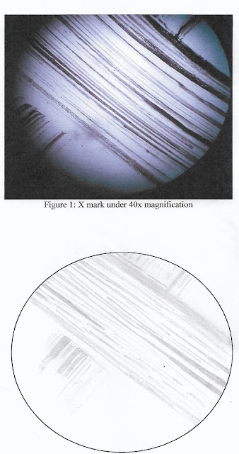
This
experiment was conducted to learn how to use a simple bright-field microscope
correctly. All part of the microscope has its own function and technique to be
used. The image of the sample that are observed from the light microscope was
actually the shadow of the object that are magnified from the sample. In this
experiment, the glass slide was marked with “x” and was observed under the
microscope under 40x, 100x and 400x magnification. In order to be able to see
the X mark, the coaxial knobs and the lamp condenser are needed to be adjusted
differently according to the magnification of the objective lens for a better
result. X mark are observed from 4x objective lens up to 40x objective lens.
The results obtained are as on the figures above and can be observed as the
magnification increase, the image of X mark can be seen clearly as it was
magnified and more detail of the X mark can be clearly observed.
CONCLUSION:
Microscope is an instrument that are used to observe an image or microorganism clearer. Based on the experiment that had been conducted, we can conclude that ‘X’ mark is clearer with 400x magnification compared to 40x & 100x magnification. The higher the magnification, the clearer the image. Therefore, the objective of this experiment is achieved.
REFRENCES
Lab
manual
1.2 Examination
of Cells
The cells can be identify by using microscope. In light microscopy, oil immersion is a technique used to increase the resolution of a microscope. This can be achieved by immersing both the specimen slide and the objective lens which this can increased the numerical aperture of the objective lens. Many condenser also give optimal resolution when the objective lens is immersed in oil. Therefore, we using microscope to observe e-coli which is very small in size.
INTRODUCTION:
The cells can be identify by using microscope. In light microscopy, oil immersion is a technique used to increase the resolution of a microscope. This can be achieved by immersing both the specimen slide and the objective lens which this can increased the numerical aperture of the objective lens. Many condenser also give optimal resolution when the objective lens is immersed in oil. Therefore, we using microscope to observe e-coli which is very small in size.
The other method that can be used is called wet
amount method. Wet amount is a glass slide holding a specimen suspended in a
drop of liquid for microscopic examination. Wet amount are necessary to
examining living organism under microscope. This method also allows the
observer to watch the motility of the microorganism.
OBJECTIVE
- To gain the knowledge and experience in the use of microscope
- To enhance the skilfulness in the use of microscope
- To observe the diversity of cell and microorganisms.
MATERIALS AND REAGENTS
- Lens tissue
- Slide and cover slip
- Immersion oil
- Culture (E.coli)
- A microscope slide containing stained microorganisms
- Bunsen burner
- Inoculating loop
PROCEDURE
Stained
cells:
- Microscope was set up based on the procedure given and the slide was examined under the oil immersion lens.
- The shape and the size of the organisms also including the structure that was found through the observation had been taken using a camera.
The
wet mount:
- A sterile Pasteur pipette was used to aseptically transfer one drop of culture (E.coli) to the centre of a glass slide.
- A mark was drawn on a coverslip by using a marker pen to help us more focus on the microorganisms.
- The coverslip was taken and was turned to ensure the marker pen faced down. Then, one edge of the coverslip was placed and lowered gently onto the slide to cover the drop of culture. The culture was spread in between the coverslip and the slide.
- The slide was placed on the microscope stage and the 4x objective lens was set up to focus the culture.
- The 10x and the 40x objective lens were used in observing the cell. Smaller moving objects were detected randomly within certain times when observed more closely. Even some of the bacteria observed were larger in size.
- The cells was observed using oil immersion lens. At this time, the condenser and diaphragm were reconfigured.
RESULT

Figure 1.2: E.coli
under 40x magnification

Figure 1.3:
E.coli under 40x magnification

Figure 1.3:
E.coli under 100x magnification

Figure 1.4:
E.coli under 100x magnification
DISCUSSION
When light passes form a material of one refractive index to another, its bends. Light is reflected and scatters in the space between the microscope objective lens (air, refractive index, 1.0) and the slide (glass, refractive index 1.5). When light passes through both glass and air, it is refracted. Light of different wavelengths bends at different angles. When using lower magnification microscope objective lenses (4x, 10x, 40x), the light refraction is usually not noticeable. However, under high magnification microscope objective lens (100x), light refracts in different ways, this causes the image to be unfocused. In microscopy, clearer and crisper images have to be produced in higher intensity of light. To reduce the amount of light refraction, light have to direct through a very narrow diameter of a higher power objective lens. Immersion oil is substance with a refractive index equal to that of the glass slide which can direct more light through the objective lens. This increases the resolution of the image due to greater light-focusing ability and reduces light scattering as light passes from the specimen to the objective lens. Therefore, a clearer image is observed.
When light passes form a material of one refractive index to another, its bends. Light is reflected and scatters in the space between the microscope objective lens (air, refractive index, 1.0) and the slide (glass, refractive index 1.5). When light passes through both glass and air, it is refracted. Light of different wavelengths bends at different angles. When using lower magnification microscope objective lenses (4x, 10x, 40x), the light refraction is usually not noticeable. However, under high magnification microscope objective lens (100x), light refracts in different ways, this causes the image to be unfocused. In microscopy, clearer and crisper images have to be produced in higher intensity of light. To reduce the amount of light refraction, light have to direct through a very narrow diameter of a higher power objective lens. Immersion oil is substance with a refractive index equal to that of the glass slide which can direct more light through the objective lens. This increases the resolution of the image due to greater light-focusing ability and reduces light scattering as light passes from the specimen to the objective lens. Therefore, a clearer image is observed.
The
microorganism, Escherichia coli is
observed using oil immersion technique. Escherichia
coli is rod in shape, very tiny in size. From the observation, there are
too many E.coli there because the
culture used are concentrated. The size or the shape can be seen clearly as it
clump together or too close to one another. The result obtained, cannot be seen
clearly as the bacteria is colourless as the culture was not added with dye to
cause colour changes. In order for a better observation for this experiment,
the culture concentration need to be dilute and dye need to be added for a
better observation.
In
conclusion, the objective of this experiment which is to gain the knowledge and
experience in the use of microscope and enhance the skilfulness in the use of
microscope and to observe the diversity of cell and microorganisms is achieved.
As E.coli are successfully been
observed under the light microscope.
REFERENCE
https://www.ncbi.nlm.nih.gov/pmc/articles/PMC2134870/

Comments
Post a Comment