LAB 5: DETERMINATION OF ANTIMICROBIAL EFFECTS OF MICROBIAL EXTRACTS

SCHOOL OF TECHNOLOGY
INDUSTRY
UNIVERSITY SCIENCES
MALAYSIA
DEGREE IN BIOPROCESS
IBG 102
BIOLOGY FOR
TECHNOLOGIST
Name
|
1.
LOH SHI WEI (137602)
2.
LAI CHONG SING (137592)
3. SITI NORASYIKIN BINTI
SALMI (137673)
4. SITI NUR SUHAILI AFIQAH
BINTI SARIMAN (137674)
5.
NUR LIYANA ATHILAH BINTI
MOHD AFFANDI (137636)
|
Title
|
LAB 5: DETERMINATION OF ANTIMICROBIAL
EFFECTS OF MICROBIAL EXTRACTS
|
Date of practical
|
10/10/17
|
Date of Report Submitted
|
24/10/17
|
Lecturer
|
DR MOHD NAZRI ISMAIL |
LAB 5: DETERMINATION OF ANTIMICROBIAL EFFECTS OF
MICROBIAL EXTRACTS
OBJECTIVE
To determine the antimicrobial effects of extracellular extracts of selected LAB strains
INTRODUCTION
Some group of certain bacteria has the ability to
produce antimicrobial substances that capable to inhibit the growth of
pathogenic and spoilage microorganism. Antimicrobial substances is a substances
that harmful to microorganism by either killing or inhibiting growth. “Agents
that actually kill organisms are called –cidal
agents, with a prefix indicating the type of microorganism killed. Thus, bactericidal,
fungicidal, and viricidal agent kill bacteria, fungi and viruses respectively.
Agents that do not kill but only inhibit growth are called –static agents, and include bacteriostatic, fungistatic and
viristatic compounds” (Michael 200).
Bacteriocins are a kind of ribosomal synthesized
antimicrobial peptides produced by bacteria, which has ability to kill or
inhibit bacterial strains closely-related or non-related to produced bacteria
but will not harm bacteria themselves by specific immunity proteins. Bacteriocin
can be produce by either gram positive or gram negative bacteria. Among gram
positive bacteria, bacteriocin produce by lactic acid bacteria (LAB) have
gained particular attention nowadays. These substances can be used in the food
industry as natural preservatives. The use of LAB and their metabolic product
is generally considered as safe. The application of the produced antimicrobial
compounds as a natural barrier against pathogens and food spoilage caused by
bacterial agents has been proven to be efficient. Different classes of LAB
bacteriocins have been identified on the basis of biochemical and generic
characterization. These bacteriocins have been reported to inhibit growth of Listeria monocytogenes and, S.aureus.
MATERIALS AND APPARATUS
MRS
broth
Sterile filter paper disk (50 mm x 50 mm)
Sterile filter paper disk (50 mm x 50 mm)
Forceps
Sterile universal bottle
Cultures of LAB and spoilage/ pathogenic organism
Bench-top refrigerated centrifuged
Incubator 30°C and 37°C
UV/V is spectrophotometer
Distilled deionized water
Trypticase soy agar
Brain heart infusion agar
Yeast extract
Sterile universal bottle
Cultures of LAB and spoilage/ pathogenic organism
Bench-top refrigerated centrifuged
Incubator 30°C and 37°C
UV/V is spectrophotometer
Distilled deionized water
Trypticase soy agar
Brain heart infusion agar
Yeast extract
PROCEDURE
Part 1:
Determination of bacteriocin activity via agar diffusion test.
1. All the petri
dishes according to the spoilage organisms and strains of LAB used are labelled.
2.
Each plate will be used for one strain of
spoilage organism and one strain of LAB. The plate is divided into 2, each side
for one replicate.
3.
Each group
will have 3 strains of LAB and 3 trains of spoilage/pathogenic organisms.
4.
10 ml of
tryticase soy-yeast extract agar (TSAYE) is loaded into the labelled petri
dishes and the agar is ensured to cover the entire
surface of the plate. The agar is waited until it became solidified.
5.
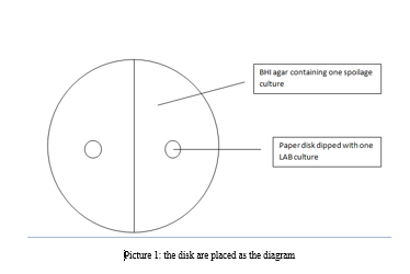
2 ml of the
broth containing the spoilage organism is inoculated into 10 ml of the brain
hear infusion (BHI) agar and then it is being vortex.
6.
The mixture is
loaded on the top of the TSAYE agar layer and is ensured that it covers the
entire surface. The gar is waited until it became solidified.
7.
The broth
containing LAB cultures is centrifuged. The supernatant will be used as
extracellular extracts.
8.
A sterile filter
paper disk is aseptically picked up with sterile forceps and is dipped into the
extracellular extract. The excess extract is ensured to have drained off.
9.
The paper disk is
placed on the top of the solidified BHI agar.
10. Next, the plates is incubated for 24 – 28 hours at
37˚C.
11. Upon incubation, the inhibition zones is measured in
cm and the readings is recorded.
Part 2: Determination
of bacteriocin activity via optical density.
1.
The broth
containing LAB cultures is centrifuged. The supernatant will be used as
extracellular extract.
2.
Each group will
have 3 strains of LAB and 3 strains of spoilage/pathogenic organism.
3.
5 ml of
double-strength MRS is added with 1 ml of cultures containing
spoilage/pathogenic bacteria and vortex the mixture.
4.
A serial
dilution of extracellular extracts (diluted 0x, 2x, 10x, 50x, 100x) is
prepared.
5.
5 ml of each
extracellular extract dilution is added into the mixture as prepared in step 3.
6.
The mixture is
incubated for 12-15 hr at 37°C.
7.
A control is
prepared using 5 ml of double strength MRS, 1 ml of cultures containing
spoilage/pathogenic bacteria and 5 ml of sterile peptone. The mixture is
incubated for 12-15 hr at 37°C.
8.
A
negative-control is prepared for 'auto-zero' via the spectrophotometer 5 ml of
double strength is added with 2 ml of distilled water ( Need not incubate)
9.
Upon incubation,
the optical density of the spoilage/ pathogenic bacteria is measured at 600 nm.
Performed the same for the control as well.
10. One arbitrary unit (AU) is defined as the dilution
factor of the extracellular extract that inhibit 50% of the spoilage/pathogenic
bacteria growth and expressed as AU/ml.
11. 50% of the spoilage/pathogenic bacteria growth are
determined from the OD600 of the control.
RESULTS


DISCUSSION
Optical density measurements can be used to determine the biomass
concentration in a suspension, when, for instance,
monitoring the growth of a culture of microorganism.
When visible light passes through a cell suspension, the light is scattered.
Greater degree of scatter indicates that more bacteria are present.
A
spectrophotometer can be set at a wavelength of 420 – 660 nm. In this
experiment, the OD600 is measured. OD600 is an abbreviation indicating the
optical density of a sample is measured at a wavelength of 600 nm. Different
vegetative cells and bacterial spores may not have the same maximal absorbance
wavelength. OD600 is chosen because at this wavelength, the cells will not
be killed due to the exposure of too much light intensity.
The control to show the growth of
bacteria without extracellular extract of lactic acid bacteria has been set up
for the pathogenic bacteria. TheOD600 of the control was then
measured in order for us to investigate whether there is inhibition of pathogenic
bacteria activity by comparing the OD600 of the samples. If the OD600
of the sample is less than OD600 of the control, there will be inhibition
of spoilage bacteria.
One
arbitrary (AU) is known as the dilution factor of the extracellular extract
that inhibited 50% of the spoilage or pathogenic bacteria growth and expressed
as AU/ml.
Control: Abs600 = Z. Thus, 50% of Z = Z/2
y = mx + c; Thus, x = (y-c)/m
When y = Z/2, Thus x = (Z/2 -c)/m
The
lower the concentration of extracellular extract, the lower concentration of
bacteriocin, the higher the growth rate of bacteria.
For
the graph plotted from the data of LAB 1:
L. casei shirota (Yakult), a negative gradient from 0x to 2x dilution,
which means LAB 1 does not clearly show inhibition on Staphylococcus. From 2x
to 10x, a positive gradient graph is obtain shows LAB 1 inhibit the growth of Staphylococcus. From 10x to 100x, a
negative gradient graph is obtain means LAB 1 does not clearly show inhibition
on Staphylococcus.
For the graph plotted from the data
of LAB LAB 2: L. sakei a positive
gradient from 0x to 10x dilution, indicates LAB 2 shows positive inhibition on Staphylococcus. However, negative
gradient is obtained from 10x to 100x LAB 2 does not clearly show inhibition on
Staphylococcus.
For
LAB 1, there is a decreasing trend on the growth of bacteria from 0x to 2x and
10x to 100x dilution. For LAB 2, there is a decreasing trend on the growth of
bacteria from 10x to 100x dilution. The result obtained might not be accurate
due to the improper preparation of the serial dilution solution. This causes
the result obtained is not like what it is supposed to be. This means that both
graph should always have positive gradient from 0x to 100x dilution
CONCLUSION
In conclusion, the objective of the experiment which is to determine the antimicrobial effects of extracellular extracts of selected LAB strains is success.
REFERENCES
In conclusion, the objective of the experiment which is to determine the antimicrobial effects of extracellular extracts of selected LAB strains is success.
REFERENCES
WEB
https://academic.oup.com/jac/article/6/4/424/727780/Bacteriocins-are-they-broad-spectrum-antibiotics
BOOK
Madigan, M. T., Martinko, J. M., Bender, K. S.,
Buckley, D. H., & Stahl, D. A. (2015). Brock biology of microorganisms (Fourteenth
edition.). Boston: Pearson
Comments
Post a Comment